Ultra-Wide Field Retinal Imaging Device
Realizing retinal imaging diagnostics with a reduced burden on patients
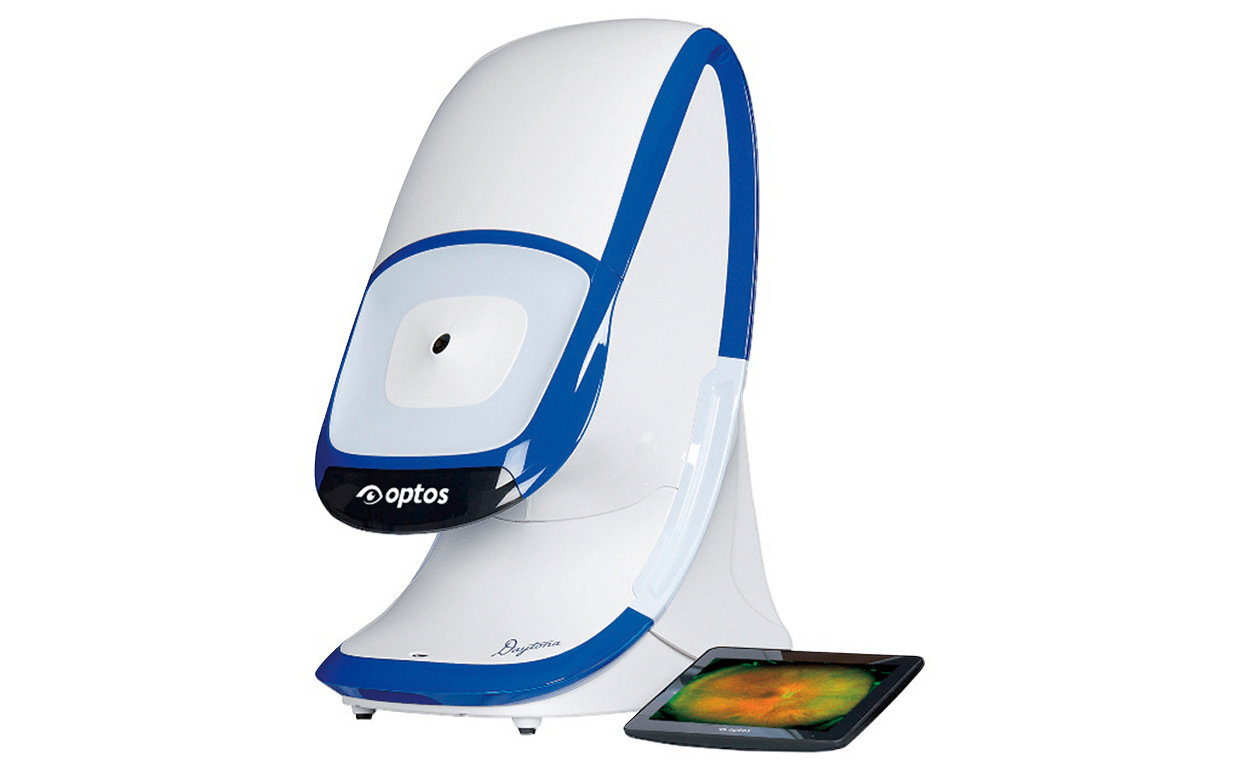
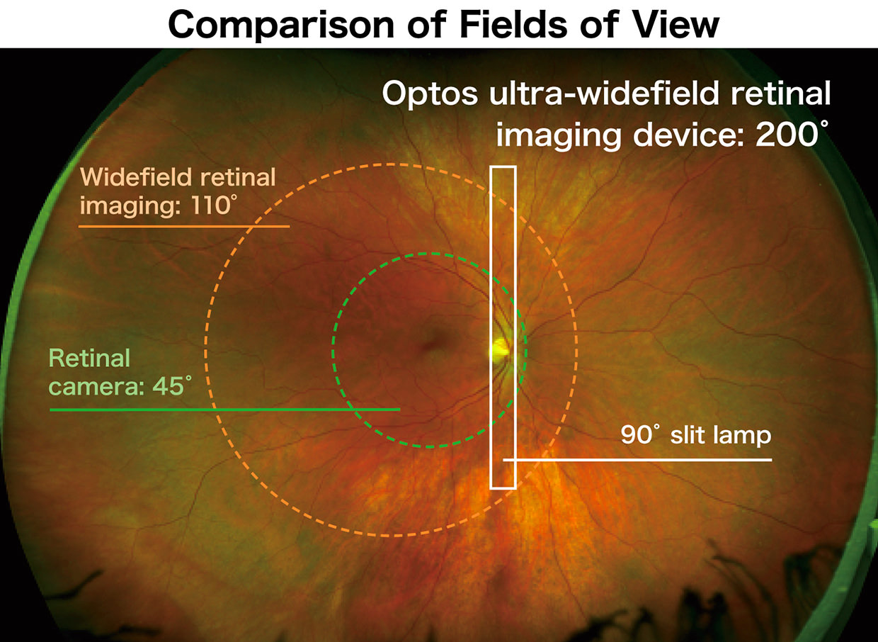
Nikon group's Optos has several ultra-widefield high-resolution imaging equipments. Its ultra-wide field retinal imaging device provides simultaneous non-contact pole-to-periphery views of approximately 80% or 200° of the retina in a single, patient-friendly capture within 0.4 second. As well as detecting eye problems such as a retinal detachment, it is used for diagnosis of systemic disease such as diabetes due to diabetic retinopathy, so is effectively utilized at ophthalmology medical facilities.
With conventional retinal imaging equipment it is necessary to capture images repeatedly and join them together to obtain the same 200° picture angle as Optos equipment, which requires a lot of time. Optos retinal diagnostic imaging equipment provides non-mydriatic diagnosis, so the waiting time of approximately 30 minutes to 1 hour after applying instillation is eliminated. Also, continuous diagnosis photography is available, which greatly shortens inspection time. Because of non-mydriatic diagnosis, patients receive various benefits such as allowing them to return to everyday life right after inspection. Utilizing different wavelengths of laser light reduces the risk of missing symptoms, which are difficult to observe with conventional equipment.
Optical system employing an ellipsoidal mirror
For conventional retinal cameras, a mydriatic is needed for opening pupils to photograph the retina. However, Optos ultra-wide field retinal imaging device achieves ultra-widefield photography with a wide field of view of 200° without mydriatic. This is realized by a unique optical system utilizing an ellipsoidal mirror, which has a concave three-dimensional shape, and laser light that scans the retina. Laser light reflects on the ellipsoidal mirror, and scans the retina through the patient's pupils. Irradiating laser light by changing the angle of laser incidence using a galvanometer mirror, which enables high-speed scanning by reciprocating oscillating, an ultra-widefield, high-definition image can be taken at high speed.
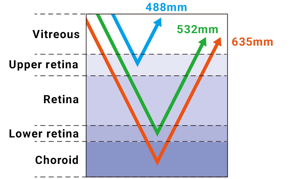
Using multiple laser lights for scanning
To realize various observations, Optos technology incorporates two kinds of scanning laser (Red 635nm; Green 532nm) that cover different distances. The green laser scans from the sensory retina to the pigment epithelial layers, while the red laser scans from the RPE*1 to the choroid. Also, high-end equipment employs a blue scanning laser for fluorescein vascular inspections*2. In this way, lesion areas can be determined easily.
- *1RPE: Retinal pigment epithelium
- *2Fluorescein vascular inspections: Fluorescein is injected into vessels mainly for examine lesions to retina and the retinal pigment epithelium.
Product Technology
- Digital SLR cameras
- Mirrorless cameras
- Binoculars
- FPD Lithography Systems
- Semiconductor Lithography Systems
- Alignment Station
- Super Resolution Microscopes
- Ultra-Wide Field Retinal Imaging Device
- Video Measuring Systems
- Industrial X-ray/CT Inspection Systems
- Non-Contact Large-Volume Inspection System