Super Resolution Microscopes
Optical microscopes redefined by groundbreaking technologies
Super resolution microscopes have been gaining in popularity on the front lines of life science research today, as they allow observation at much higher resolutions than conventional optical microscopes. Nikon's innovations realize nanometer-level observation, delivering a direct view of the most intricate structures in cells.
In the field of optics, including cameras and microscopes, resolution or resolving power is defined as the capacity to differentiate two close objects. The maximum resolution of conventional microscopes in general is usually about 200 nanometers, due to the objective lens size and light wavelength. Super resolution microscopes offer much higher resolution than this.
Realizing observation of intricate organelle structures, which used to be impossible, super resolution microscopes are expected to greatly contribute to the advancement of diverse fields, including medicine and biology, where nanometer-level observations are critical.
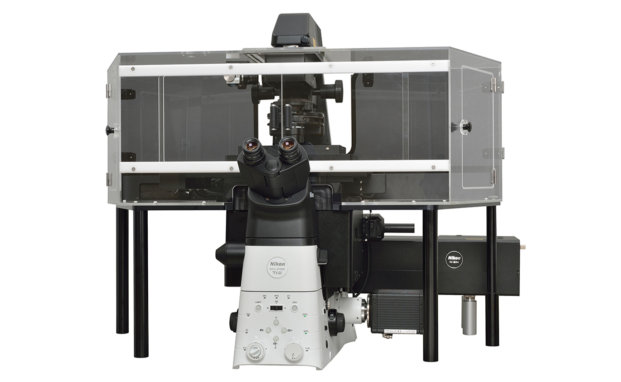
N-SIM
The Super Resolution Microscope N-SIM series attains spectacularly high resolution of nearly double that of conventional optical microscopes, by combining Nikon's unique optical technologies and Structured Illumination Microscopy (SIM), a technique announced by Dr. Mats G.L. Gustafsson*1 at the University of California, San Francisco in 2000.
When a finely striped, patterned illumination (structured illumination) is applied to a specimen, it produces moiré patterns. These patterns are characteristically coarser than the original, and can be captured by optical microscopes, even in the case of a sub-resolution specimen.
N-SIM captures multiple moiré images while slightly varying the orientation and phase of the structured illumination. Then it analytically processes those images to mathematically recreate the intricate structures of the specimen. Adopting this groundbreaking technology, N-SIM achieves significant resolution by using a new illumination scheme without drastic alteration of the microscope mechanism.
As well as this, Nikon has developed an innovative high-speed, structured illumination system. The N-SIM S Super Resolution Microscope realizes high-speed, super-resolution imaging*2 at a maximum of 15 fps (frames per second).
- *1Group leader (2008-2011), Janelia Farm Research Campus, Howard Hughes Medical Institute.
- *2When exposing 512x512 pixels with 2D-SIM mode for 2 msec.

N-STORM
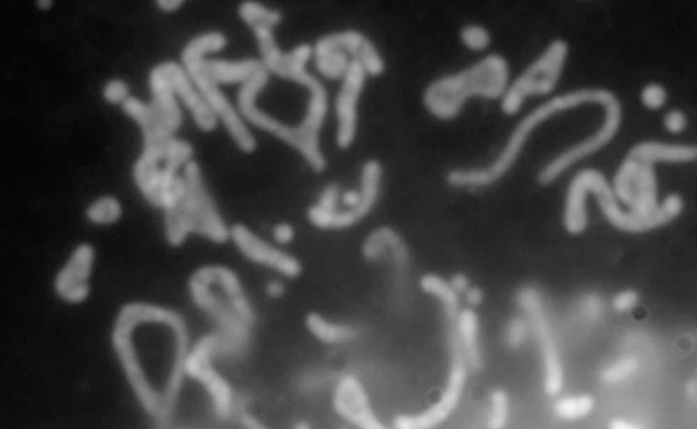
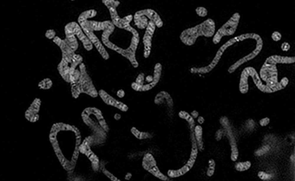
Super Resolution Microscope N-STORM for fluorescent observation is a product of collaboration with Harvard University, adopting the institution's STochastic Optical Reconstruction Microscopy (STORM) technology.
Fluorescent observation uses rays such as those of visible light irradiated onto a specimen to excite fluorophores and captures the emitted fluorescent elements as an image. However fluorophores have been known to emit light but then deteriorate in color simultaneously, without subsequent illumination. When several fluorophore molecules of an observation specimen were positioned separated by less than 200 nanometers — beyond maximum resolving power, it was impossible to distinguish and observe them as anything other than a single clump by conventional microscopes.
N-STORM uses newly discovered fluorophores, that repeatedly blink under certain conditions. The microscope irradiates laser lights onto those fluorophores to repeat the illumination, captures the elements and locates the molecules in high precision down to a few dozen nanometers. Repeating this process 5,000 to 10,000 times, the microscope creates an image of the specimen, like a stippled picture. Thanks to this new technique, N-STORM realizes resolution 10 times greater than conventional optical microscopes.

Product Technology
- Digital SLR cameras
- Mirrorless cameras
- Binoculars
- FPD Lithography Systems
- Semiconductor Lithography Systems
- Alignment Station
- Super Resolution Microscopes
- Ultra-Wide Field Retinal Imaging Device
- Video Measuring Systems
- Industrial X-ray/CT Inspection Systems
- Non-Contact Large-Volume Inspection System