A1 HD25/A1R HD25 Confocal Microscope
The world's largest, 25mm field of view confocal microscope for high-throughput, high-resolution imaging
August 30, 2018
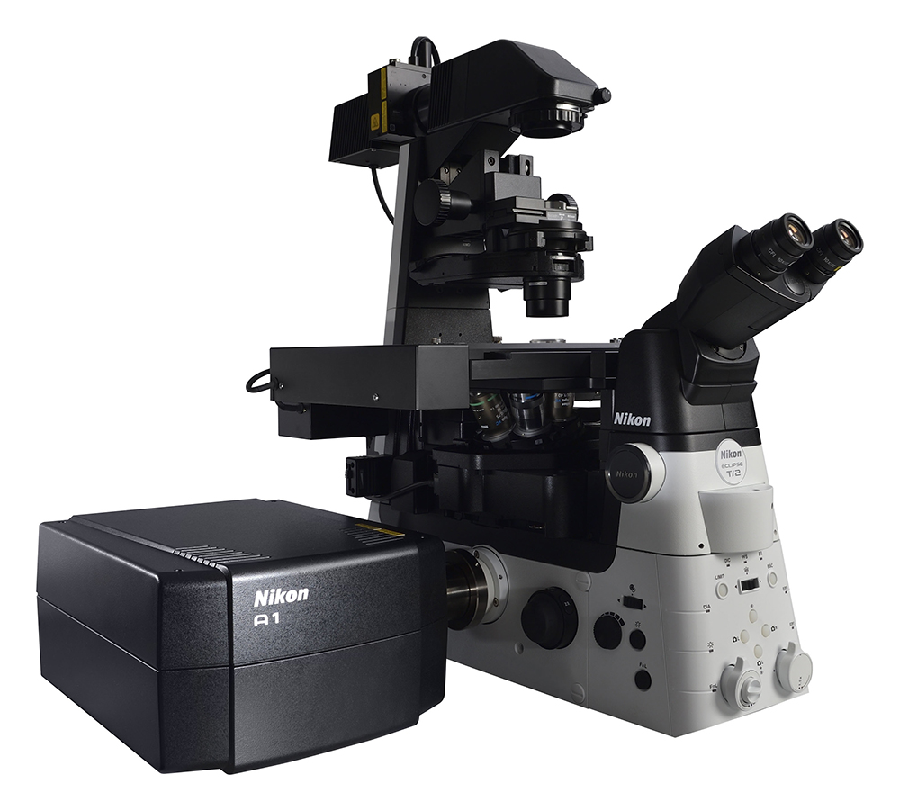
(Combination with ECLIPSE Ti2 inverted research microscope)
TOKYO - Nikon Corporation (Nikon) is pleased to announce the release of the A1 HD25/A1R HD25 confocal microscope, which enables high-speed, high-resolution image acquisition of large areas utilizing the world's largest*1 FOV*2 (25 mm), nearly twice the field of view of conventional models.
- *1As of August 30, 2018, among point scanners for sale, according to research conducted by Nikon.
- *2Field of view: the diameter of the observable image in a microscope.
Product Information
| Product Name | A1 HD25 confocal microscope A1R HD25 confocal microscope |
|---|---|
| Availability | August 31, 2018 |
Product Concept
A confocal microscope is an optical sectioning system that enables acquisition of high-contrast, high-resolution images by eliminating out-of-focus light using a pinhole placed on the detection side. These optical sections can be acquired at different heights within a sample and combined to generate 3D images of thick samples.
There is an ever-increasing demand in bioscience research for observation of large samples such as tissues and whole organisms, and dynamic events occurring within them.
In response to this demand, Nikon has developed the A1 HD25/A1R HD25 confocal microscope, which achieves the world's largest FOV confocal imaging. By capturing more data in a single image, Nikon's new confocal can reduce acquisition times without sacrificing resolution or magnification. Combined with the high-definition resonant scanner, the new confocal system enables high-speed, high-throughput imaging of live, whole organisms such as zebrafish and drosophila.
Main Features
1. Large image acquisition with the world's largest FOV
In combination with the ECLIPSE Ti2-E inverted research microscope, the A1 HD25/A1R HD25 achieves the world's largest confocal FOV of 25 mm. It acquires nearly twice the area compared to conventional, 18mm FOV confocal systems, increasing both data throughput and enabling users to capture significant events occurring outside the normal field of view.
Image comparison between conventional model and new A1 HD25
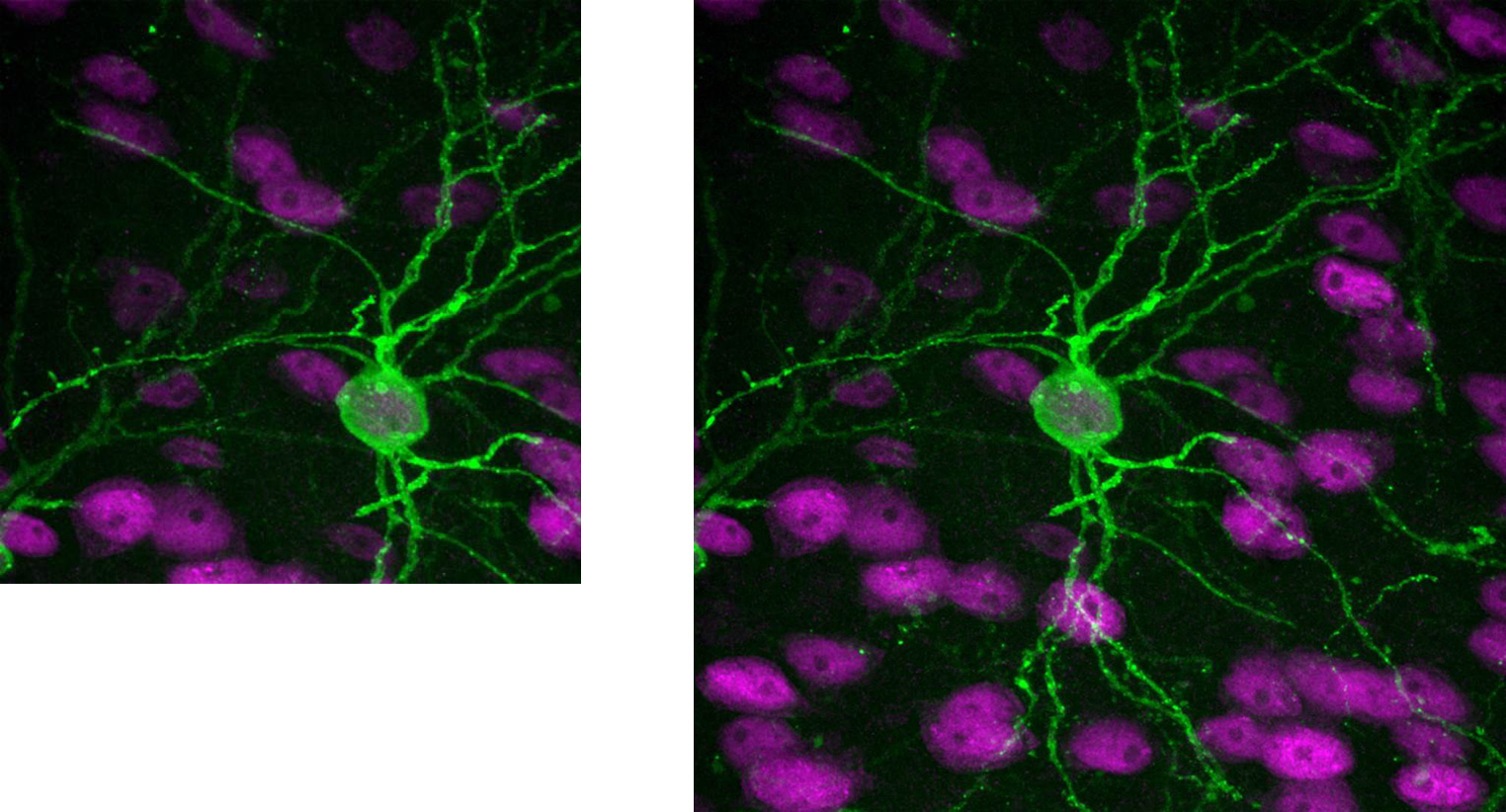
Sample: Cranial nerve of marmoset brain
2. High-speed imaging of up to 720 fps to capture fast events
The A1R HD25 incorporates a high-definition resonant scanner that realizes an imaging speed of 720 fps (512 x 16 pixels). It enables acquisition of ultra-fast events such as ion signaling and also reduces photo-damage to cells and photobleaching by minimizing light exposure.
3. Double the throughput
With twice the amount of data acquired per image, the A1 HD25/A1R HD25 significantly reduces image capture times for high-throughput screening assays and large image stitching*3 applications without sacrificing resolution or magnification. Furthermore, the fast high-definition (1K, 1024 x 1024 pixels) scanner enables high-resolution, high-quality imaging even at lower magnifications, enabling observation of minute structures within samples.
- *3An image processing method that combines neighboring images to create one large image.
Specifications
![]() Swipe horizontally to view full table.
Swipe horizontally to view full table.
| A1 HD25 | A1R HD25 | ||
| Scanning area | 25 mm (square inscribed in a 25 mm-diameter circle) | ||
| Image size | Galvano scanner | Max. 4096 x 4096 pixels | |
| Resonant scanner | - | Max. 1024 x 1024 pixels | |
| Scanning speed (imaging speed) | Galvano scanner | 10 fps (512 x 512 pixels, bi-directional) 200 fps (512 x 16 pixels, bi-directional) |
|
| Resonant scanner | - | 30 fps (512 x 512 pixels) 720 fps (512 x 16 pixels) |
|
The information is current as of the date of publication. It is subject to change without notice.