“RetinaStation”
Fundus Camera with Fully Automatic, Intuitive Operation
Oct. 9, 2019
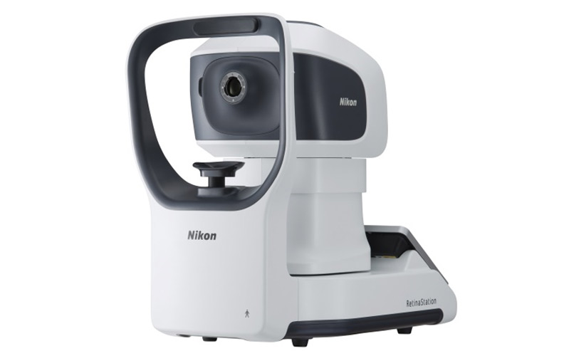
TOKYO - Nikon Corporation (Nikon) is pleased to announce the release of “RetinaStation”. The camera is able to capture high definition fundus images with full automation.
Overview of Release
| Name of Item | Fundus Camera: “RetinaStation” |
|---|---|
| Release Date | From the start of October, 2019 |
This non-mydriatic fundus camera detects abnormal changes in the retina at the back of the eye, without the use of a mydriatic agent to open the pupil. The camera has been designed for use in ophthalmic and health diagnosis.
Diseases such as glaucoma, pigmentary degeneration of the retina, diabetic retinopathy, and age-related macular degeneration, are often the cause of sight loss. These conditions are becoming more common due to an aging society and changing lifestyles. The easy-to-operate fundus camera “RetinaStation”, released by Nikon for clinics and medical diagnosis facilities, has fully automatic intuitive functions which reduce the burden on both patients and medical personnel.
Our company aims to detect disease early by continuing to expand our product line, starting with “RetinaStation”. Our aim is to improve global Quality of Life (QOL) and work toward achieving a world without sight loss, so we contribute to early detection and early treatment of eye diseases while developing innovative products and utilizing AI to provide new technologies and solutions*.
- *Commercialization of AI utilized solutions are being considered for US and European markets only.
Major Features
1. Capable of Intuitive Operation with Full Auto
When Full Auto is engaged or the Alignment mode function is used, all actions, including alignment, focusing and shooting, are performed automatically. Intuitive operation is possible through the use of the large 10.1-inch touch panel.
2. Images in High Resolution
Fundus details are reproduced in high resolution with 12 million pixels, thus enabling better diagnosis.
An image, exceeding 75-degrees is easily produced with image stitching through the use of the montage function.
3. Saving Space through Compact Housing
The compact table-top device can be placed in many areas (in a practice). Since it has an integrated PC , the same unit is used to capture and edit images, which are displayed on the LCD monitor instantly. This workflow helps to save examination time.
Major Specifications
![]() Swipe horizontally to view full table.
Swipe horizontally to view full table.
| Function | Value/ Type | Remarks |
|---|---|---|
| Fundus Image | — | Non-mydriatic, Color Image |
| Field of view | >=45 degrees | |
| Minimum pupil size | 4 mm | |
| Image sensor | CMOS 12 megapixels | |
| Alignment mode | Full Auto / Semi Auto / Activate manual mode assist | Full Auto: automatically adjusts eye position and switches to the other eye Semi Auto: automatically adjusts eye position, but target eye should be manually switched by user Activate manual mode assist: user both manually adjusts eye position and switches to the other eye |
| Display | 10.1” LCD Monitor, touch Panel | |
| Dimensions | Depth: 495 mm Width: 288 mm Height: 495 mm |
The information is current as of the date of publication. It is subject to change without notice.