Nikon introduces the AX R MP multiphoton confocal microscope
High speed acquisition of high resolution, large field of view images deep within living organisms
December 1, 2021
TOKYO - Nikon Corporation (Nikon) is pleased to announce the release of the AX R MP multiphoton confocal microscope, which can acquire high resolution, large field of view images deep within living organisms at high speed.
Release Overview
![]() Swipe horizontally to view full table.
Swipe horizontally to view full table.
| Product Name | AX R MP multiphoton confocal microscope |
|---|---|
| Release Date | Spring, 2022 |
Development Background
In recent years, in research fields such as neuroscience and immunology, research regarding fast biological dynamics (e.g., cranial nerve signaling, blood flow) has been characterized by observing large samples such as live tissue and even whole animals with a microscope. These methods are continuously advancing, and as a result, there is a demand for systems that support a broad range of experiments and enable lateral and oblique observation of samples without changing their orientation.
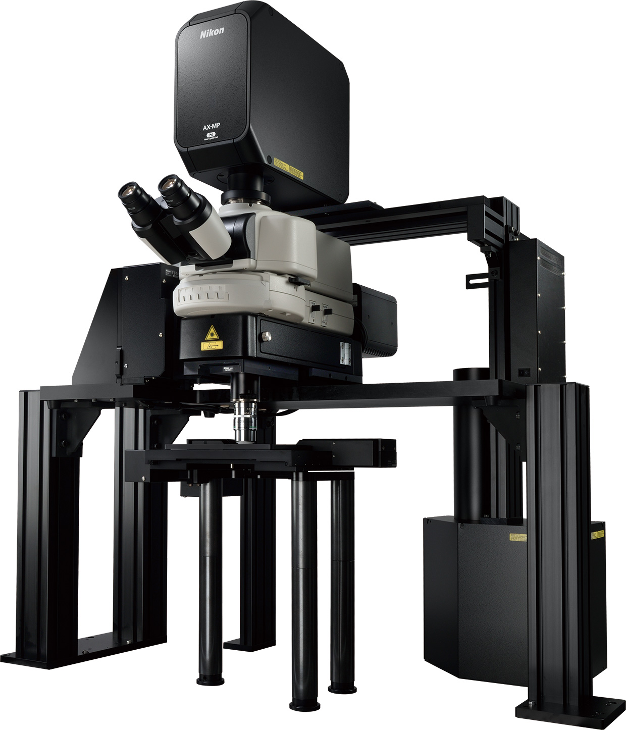
To meet such needs, Nikon manufactures high-speed multiphoton confocal microscopes that scan samples with an infrared ultrashort pulse laser*1, enabling observation of deep structures in three dimensions. And now, Nikon has developed the AX R MP multiphoton confocal microscope, which enables high speed acquisition of high resolution, large field of view images. With an upright microscope configuration, a large space is provided under the objective to support a wide range of experiments and acquisition of images from various angles. The AX R MP efficiently acquires large amounts of information on ultrastructure in the deep regions of living organisms, supporting research into diverse biological phenomena.
- *1An infrared laser with pulse width from a few femtoseconds (1/1000 trillion seconds) to a few picoseconds (1 trillionth of a second). It results in less photodamage to the object.
Main Features
1. High speed scanning of high-resolution images with a large field of view to efficiently acquire large amounts of information
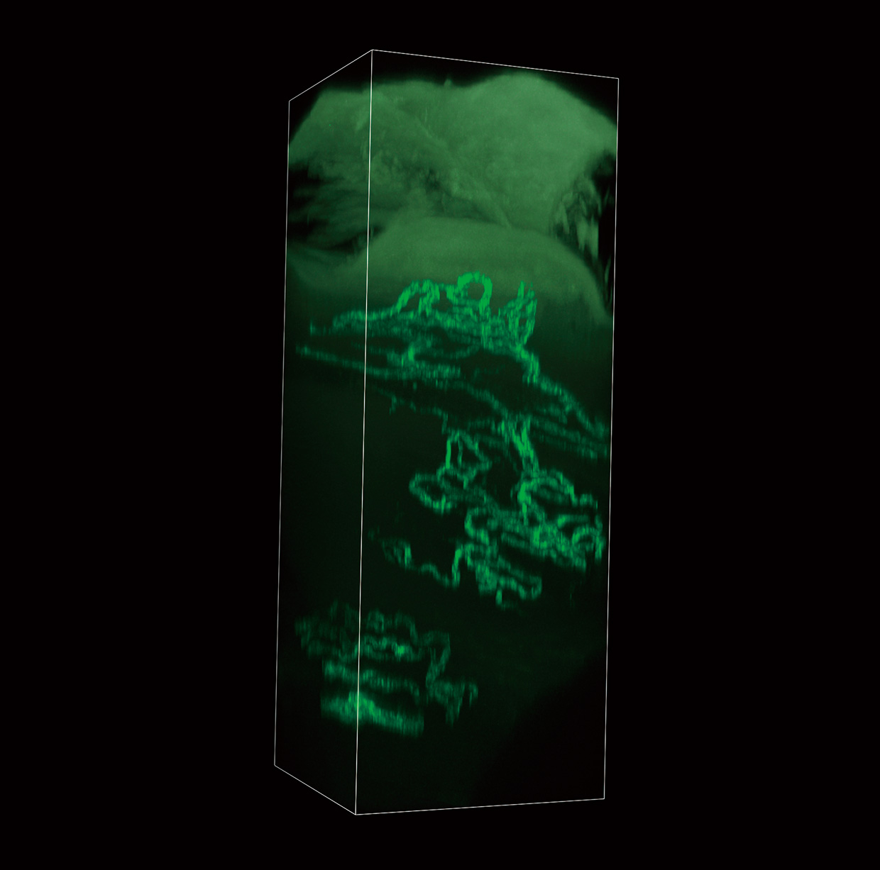
High resolution 2K x 2K (2048 x 2048 pixels) images with a large field of view*2 (22 mm) can be acquired at high speed by combining the AX R MP with the dedicated AX-FNGP and AX-FNSP microscopes for multiphoton confocal systems, or the ECLIPSE Ti2-E inverted research microscope. Fast biological activities can be captured at high resolution right up to the periphery of the field of view using a resonant scanner. Large amounts of information can be acquired efficiently, supporting research into different biological phenomena.
- *2The size of the image that can be obtained from the microscope.
The optional AX R MP-dedicated CFI75 Apochromat LWD 20XC W water dipping objective for biological microscopes was used
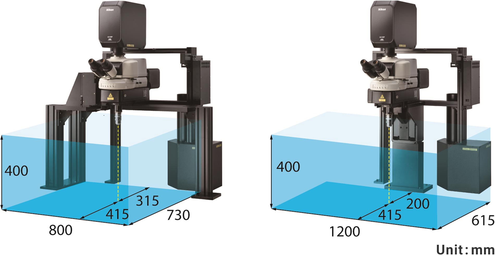
2. Responding to diverse experimental needs by providing a large space under the objective
A space with a height of 40 cm is provided under the objective of the AX-FNGP and AX-FNSP microscopes for multiphoton confocal systems to meet user needs for a sufficiently large experimental space within which large living samples can be comfortably observed. Two stands are available: a gate stand for experiments that require more depth, and a single stand for experiments that require greater width. This increases the degree of freedom when setting up samples and laboratory equipment, providing greater flexibility in adapting to large samples and different types of experiments.
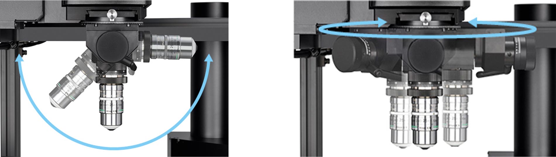
3. Achieving observation from various angles by tilting the objective
The newly developed optional CFI75 single tilting nosepiece can rotate the objective of the AX-FNGP and AX-FNSP microscopes for multiphoton confocal systems vertically or horizontally and can be adjusted to various angles. Observation may be performed without changing the orientation of the sample, resulting in an efficient workflow that saves time and effort.
4. Objectives that are capable of wide field-of-view observation of deep structures in vivo
A combination of the AX R MP with the optimum objective allows users to clearly observe the sample from the surface down to deep internal structures. High resolution objectives with chromatic aberration correction up into the near-infrared wavelength range are available to enable a variety of experimental approaches.
The optional AX R MP-dedicated CFI75 Apochromat LWD 20XC W water dipping objective for biological microscopes was released at the same time as the AX R MP. It can acquire bright images right up to the periphery of the field of view, with a design that minimizes light reduction at the periphery of the image, so that minute structures deep within living organisms can be captured with a large field of view.
The lens on the far left is the AX R MP-dedicated CFI75 Apochromat LWD 20XC W water dipping objective for biological microscopes
Specifications
![]() Swipe horizontally to view full table.
Swipe horizontally to view full table.
| Compatible microscopes | AX-FNGP microscope for multiphoton confocal systems (gate stand model) AX-FNSP microscope for multiphoton confocal systems (single stand model)
|
|---|---|
| High-speed image acquisition |
FOV 22mm resonant scanner Max. 2048 x 2048 pixels Max. 720 frames per second (512 x 16 pixels) 30 frames per second (512 x 512 pixels) |
| CFI75 single tilting nosepiece (optional) |
[Rotation] Horizontal ±100° (manual) Vertical ±90° [Z axis coarse movement] ±3 mm (manual) |
| Recommended objectives (optional) |
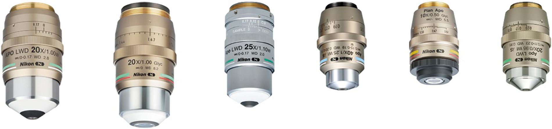
CFI75 Apochromat LWD 20XC W (NA = 1.00, WD = 2.8 mm) CFI90 20XC Glyc (NA = 1.00, WD = 8.20 mm) CFI75 Apochromat 25XC W 1300 (NA = 1.10, WD = 2.00 mm) CFI Apochromat Lambda S 40XC WI (NA = 1.25, WD = 0.20-0.16 mm) CFI Plan Apochromat 10XC Glyc (NA = 0.50, WD = 5.50 mm) CFI Apochromat LWD Lambda S 20XC WI (NA = 0.95, WD = 0.95 mm) |
The information is current as of the date of publication. It is subject to change without notice.